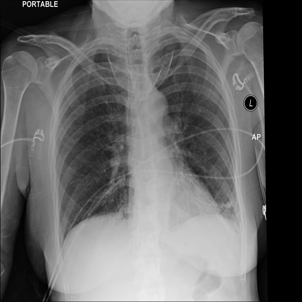
Left lower lobe consolidation, consistent with pneumonia.
Follow-up radiograph in 6-8 weeks recommended to document resolution.
Each finding, explained
ConsolidationLeft lower lobe
“Left lower lobe consolidation, consistent with pneumonia.”
Consolidation. Part of the lung where the tiny air sacs have filled with something else, usually fluid, pus or cells, so that area shows white on the image instead of dark.
Pneumonia is the most common reason. Fluid from the heart, bleeding or inflammation can look the same, which is why it's read together with symptoms.
Pneumonia. An infection in the lung, where the air sacs fill with fluid and inflammatory cells.
Treated by a clinician based on symptoms, examination and sometimes tests. A follow-up X-ray is sometimes taken weeks later to check it has cleared.
Healthy air sacs hold air (left); in consolidation they fill with fluid or cells (orange), so that part shows white on the image.