Add your radiologist's report and your scan. See what each finding means in plain words, what any grading category means, and where it is in your body.
·
Sample reading · CT CHEST WITHOUT CONTRAST · 10 February 2026
9 mm solid nodule in the right lower lobe, Lung-RADS 4A.
Mild emphysema.
Simple left renal cyst.
Each finding, explained
NoduleRight lower lobe
“9 mm solid nodule in the right lower lobe, Lung-RADS 4A.”
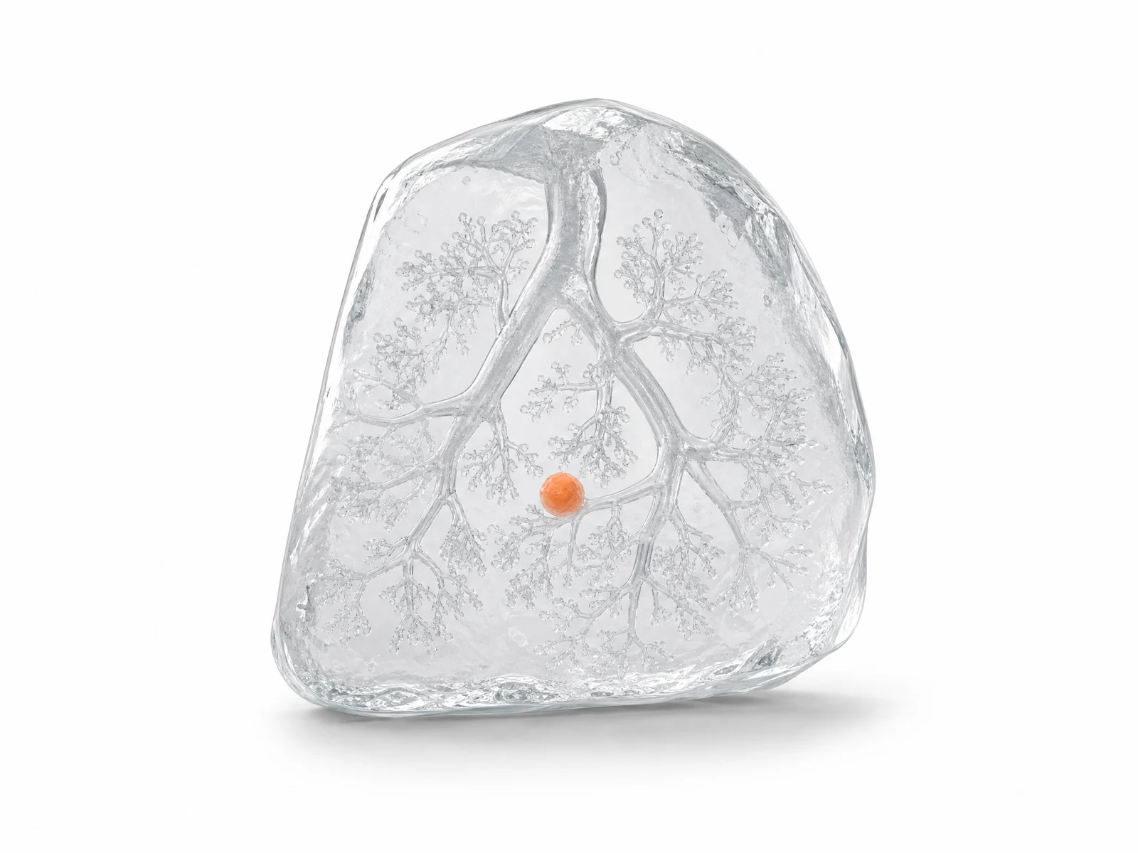
Nodule. A small, roughly round spot in the lung, up to 3 cm across.
Most are harmless, such as old scars or healed infections. Size, shape and change over time decide whether it's followed up, and the report usually says how.
A small, roughly round spot (orange) within the lung tissue.
Lung-RADS 4A · SuspiciousLung cancer screening CT scans (low-dose CT), by what the most concerning finding looks like.
1
2
3
4A
4B
4X
A finding with features that need a closer look.
The guideline pairs this with a low-dose CT in 3 months; a PET/CT may be considered when there is a larger solid part.
9 mm as measured in the report, drawn beside a pea (about 9 mm).
EmphysemaBoth lungs
“Mild emphysema.”
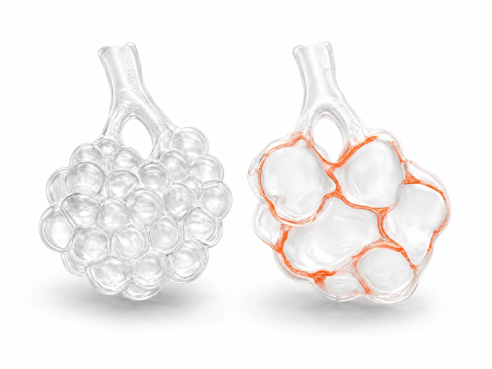
Emphysema. Some of the lung's air sacs have been damaged and merged into larger, less useful spaces.
Most often from smoking, sometimes inherited (alpha-1 antitrypsin). It's part of COPD.
Healthy small air sacs (left); in emphysema their walls break down into fewer, larger spaces (orange edges).
Kidney cystLeft kidney
“Simple left renal cyst.”
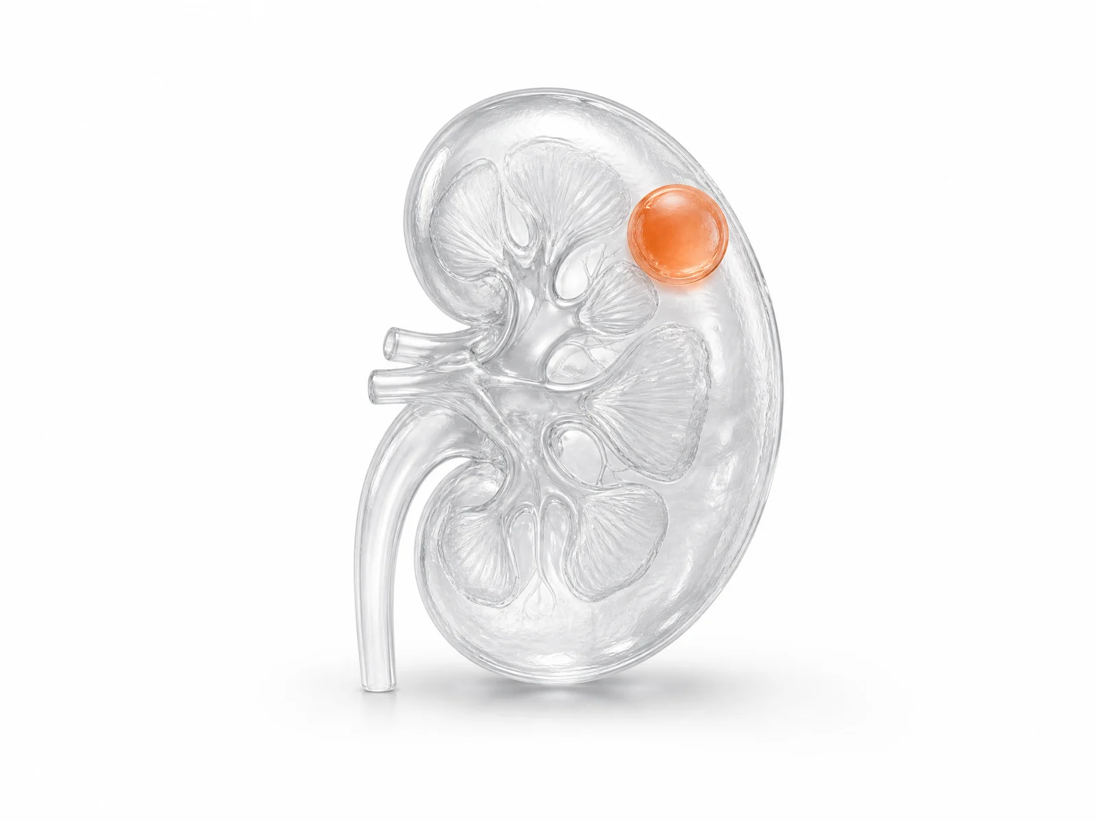
Kidney cyst. A fluid-filled sac in the kidney. A simple cyst has a thin, smooth wall and clear fluid.
Very common, especially with age, and simple ones are harmless. Cysts with more features are graded with the Bosniak classification.
A smooth, thin-walled bubble of fluid (orange) on the kidney.
Wear-and-tear in the spineUpper back (thoracic spine)
“BONES: Mild degenerative changes of the thoracic spine.”
Wear-and-tear in the spine. Age-related changes in the spine's bones and joints, such as small bony spurs.
Very common and usually noted in passing on a chest X-ray.