Sample reading · MRI BRAIN WITHOUT CONTRAST · 2 May 2026
This is how your MRI will read.
The sample brain MRISample scan: 3D Slicer sample data (MRHead). The sample report is written for this demo.
What the radiologist concluded
- No acute intracranial abnormality.
- Mild nonspecific white matter hyperintensities, commonly seen with small vessel change.
- Mild right maxillary sinus mucosal thickening.
Each finding, explained
White matter bright spotsWhite matter of the brain
“Mild nonspecific white matter hyperintensities, commonly seen with small vessel change.”
White matter bright spots. Small bright spots on T2 or FLAIR MRI in the white matter, the brain's wiring under its surface.
Common with age, high blood pressure and migraine. They have many causes, and the report sometimes suggests which is most likely.
Small vessel changes. Signs of wear in the brain's smallest blood vessels, usually seen as white matter bright spots.
Common with age, and linked to blood pressure, diabetes, cholesterol and smoking.
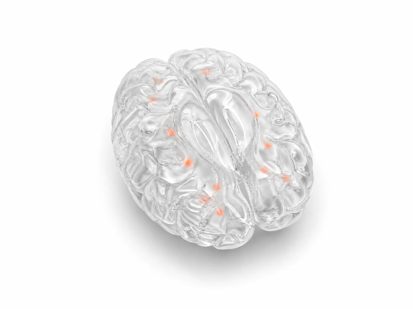 Small bright spots (orange) in the brain's white matter.
Sinus lining thickeningRight side: sinuses
“Mild right maxillary sinus mucosal thickening.”
Sinus lining thickening. The lining of one or more sinuses, the air spaces around the nose, is thicker than usual.
Very common on head scans, often from a cold, allergies or past sinus infections, and usually of no consequence.
White matter bright spotsBoth frontal lobes
“A few small foci of T2/FLAIR hyperintensity in the frontal white matter.”
White matter bright spots. Small bright spots on T2 or FLAIR MRI in the white matter, the brain's wiring under its surface.
Common with age, high blood pressure and migraine. They have many causes, and the report sometimes suggests which is most likely.
Looked at and fine
- No acute infarct.
- No mass, hemorrhage or hydrocephalus.
- The ventricles are normal in size.
Sources: RadiologyInfo.org (RSNA and ACR), Brain MRI, 2026 · RadiologyInfo.org (RSNA and ACR), glossary of radiology terms, 2026.