01Stereo depth
3D Surface Mapping
Look forNew bulges · visible asymmetry
Map the visible body surface and compare its contours. New or enlarging raised areas can be documented for examination, with their location preserved across scans.
 LifeFrom Robot v.01Back to LifeFrom
 Lie down for two minutes. It photographs your skin, watches you breathe and reads your pulse without touching you, the same way every time, so your doctor can see what changed.
Designed to refresh your vitals every day. Nothing touches you: skin, pulse and breathing are captured the same way each time, so a change is a change and not a different angle.
The instrument comes to the observation. A robotic arm carries the sensor head around you, holds steady while it records, then moves to the next view. The same routine every time, so scans line up and a change stands out.
01Stereo depth
Look forNew bulges · visible asymmetry
Map the visible body surface and compare its contours. New or enlarging raised areas can be documented for examination, with their location preserved across scans.
02Aligned depth scans
Look forLocal enlargement · changing contours
Compare the same region across 3D scans. A persistent increase in its external size can prompt a closer examination for swelling or another cause of shape change.
03Position + distance + light
Look forPersistent change across sessions
Match the viewing angle, posture and lighting before comparing scans. Help separate a repeatable change worth reviewing from a difference caused by how the image was taken.
Use steady-light photographs and video to record the face and visible skin. Compare appearance across sessions and analyse skin-colour changes for pulse estimation.
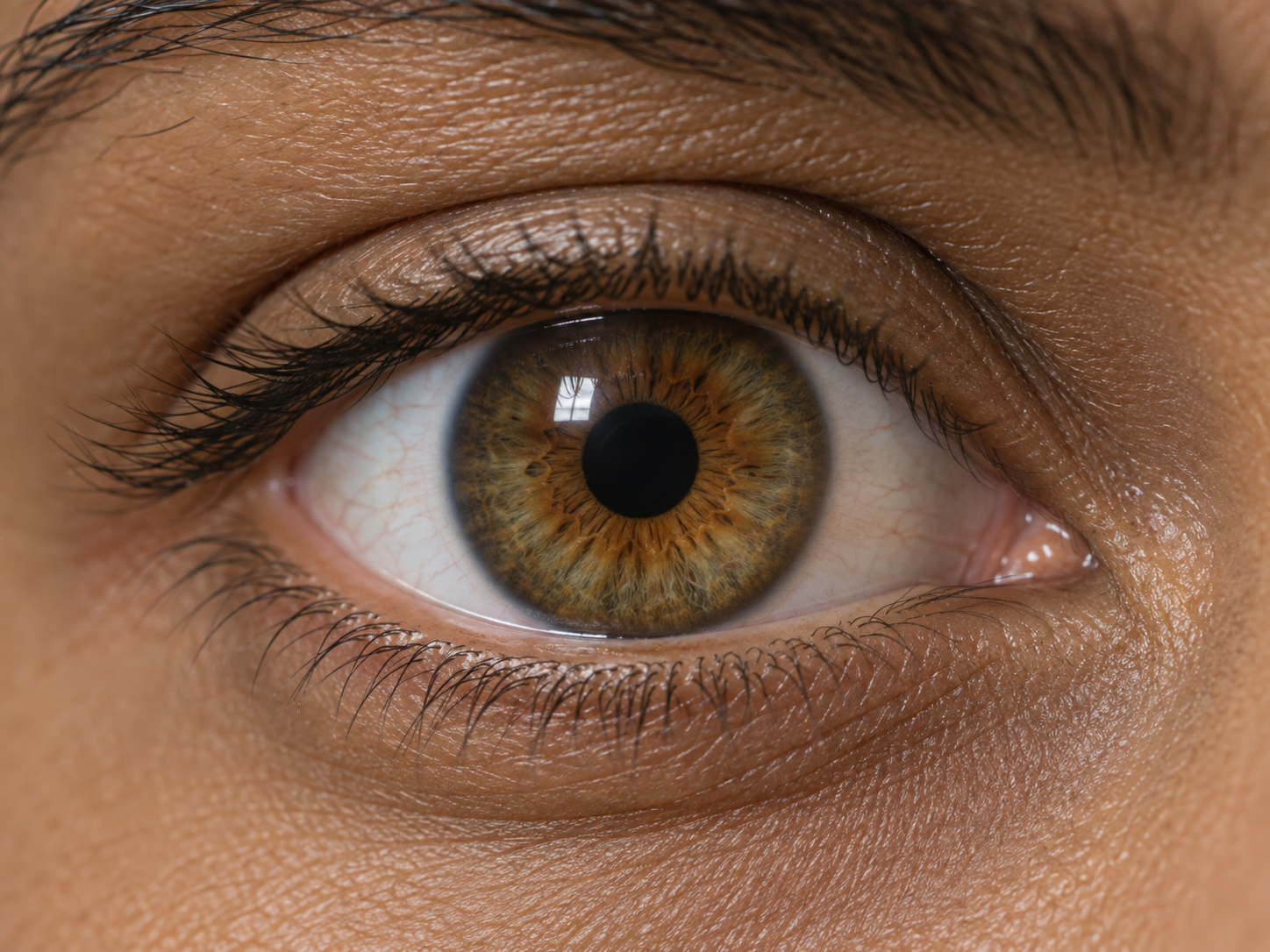
01Close-up imaging + calibrated light
Look forNew pupil asymmetry · weak light response
With sufficient eye detail and calibrated light, compare pupil size and constriction. A new size difference or reduced response can prompt an eye or neurological assessment.
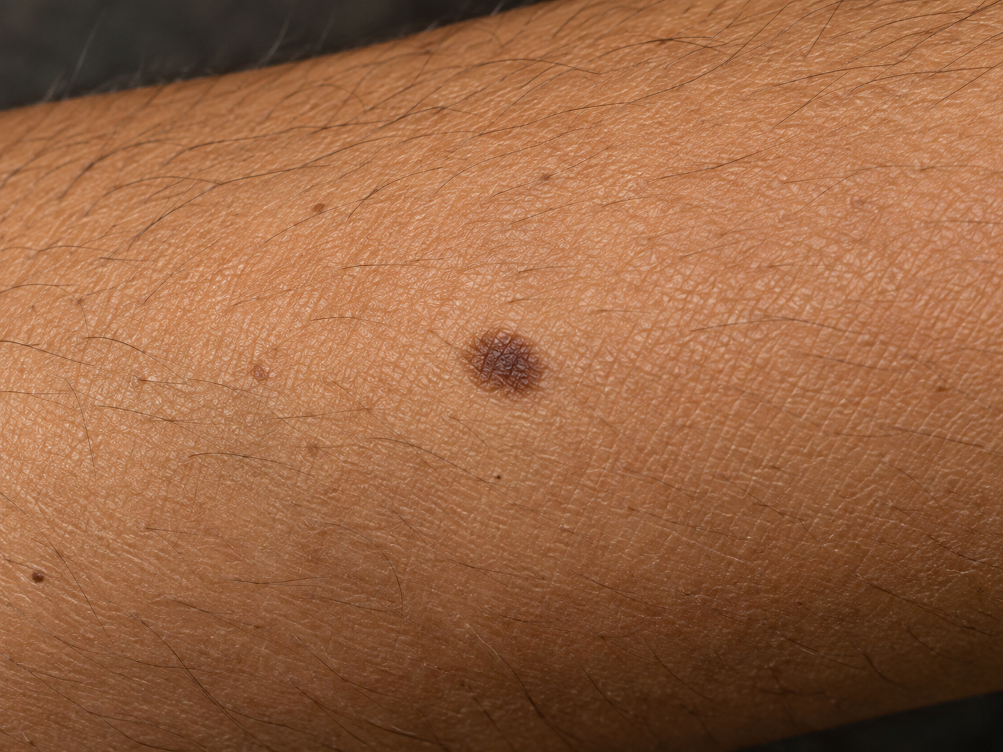
02Colour photography + depth
Look forGrowth · border or colour changes
Compare a mark’s size, borders and colour over time for dermatology review. Keep its 3D surface location; compare raised detail where the depth capture resolves it.
03RGB video analysis
Look forA resting pulse above or below your usual
Estimate pulse rate from subtle skin-colour changes in video. Persistent shifts at rest can prompt review of factors such as illness, medication or cardiovascular health.
Use successive depth frames to track the chest as it rises and falls. Analyse breathing rate, cycle timing and differences in movement between the left and right sides.
01Chest depth over time
Look forUnusually fast or slow resting breathing
Count chest-rise cycles per minute. A change in resting rate can accompany illness, pain or stress; persistent departures from your usual rate deserve review.
02Left / right depth comparison
Look forUneven expansion · reduced movement
Compare each side’s chest motion. Persistent asymmetry can suggest restricted movement—from pain, chest-wall or lung problems—and prompt a respiratory assessment.
03Breathing cycle analysis
Look forPauses in chest motion · irregular cycles
Track the timing of chest movements. Repeated pauses or uneven cycles can flag a disrupted breathing pattern to discuss with a clinician.
With the arm still, radar senses tiny movements associated with breathing and heartbeat. Its module outputs include respiratory-rate and heart-rate estimates.
0160 GHz radar · breathing motion
Look forA sustained change in breathing rate
Read breathing-related micromotion while the arm stays still. Compare the radar’s rate estimates across sessions to highlight a persistent change that may need respiratory review.
0260 GHz radar · cardiac motion
Look forA persistently high or low resting rate
Estimate heart rate from heartbeat-related micromotion. Repeated changes from your usual resting rate give you a specific trend to discuss with your doctor.
03Multi-view colour + depth fusion
Look forChanges in visible shape and skin appearance
Combine overlapping colour and depth views across guided body positions into a textured 3D map. Revisit the same area, compare its shape and appearance over time, and share the supporting captures with your doctor.
The arm returns to park. Keep images, surface maps and signal estimates together, ready to compare with earlier sessions and review changes in the LifeFrom app.
Your body. Your history. In LifeFrom.
A body map that connects every observation to you.
A face scan. A chest recording. A skin mark you want to follow. Each becomes part of your personal record, attached to the area captured and the day it was observed.
Return to the same place on your double, compare earlier scans and understand what changed. Bring that history into the conversation with your doctor.
See how a change becomes a result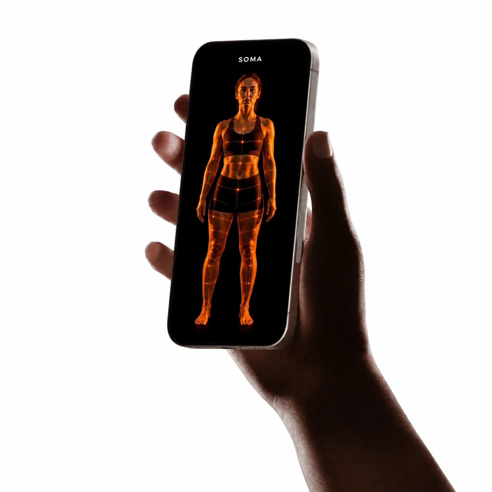
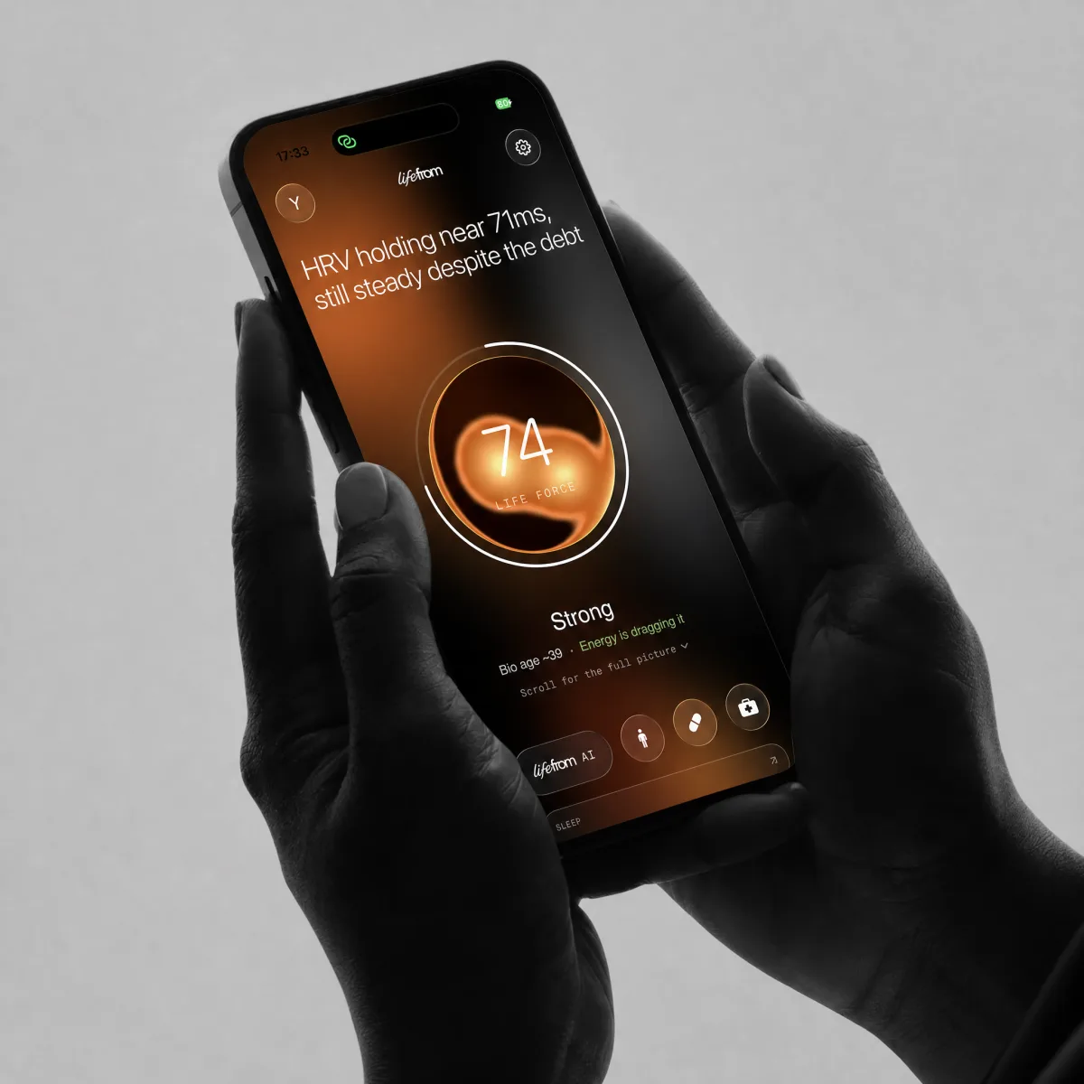
Your LifeFrom record
Understand the change. Take the next step.
Your scan becomes a clear personal update: what changed, why it may matter and what to do next. LifeFrom brings the explanation and supporting captures together, ready to share with your doctor.
“Should I get this checked?”
Ask about the finding, understand the suggested next step and prepare for your medical appointment.
Explore a resultFrom a scan to a next step
A clear explanation of what changed, why it may matter and what to do next. The supporting images and recordings stay attached, ready for a conversation with your doctor.
Explore an example app result
Send your doctor the explanation, the history and the scans behind it. Give them the evidence to decide what needs checking.

Worth a closer look
Your chest appears to move less evenly than in your earlier resting scans.
A new difference between the two sides is worth checking. Your doctor can assess whether posture, discomfort or a breathing problem could explain it.
Arrange a medical review if this change is new or persists. Bring the chest recordings and earlier comparisons to help explain what changed.